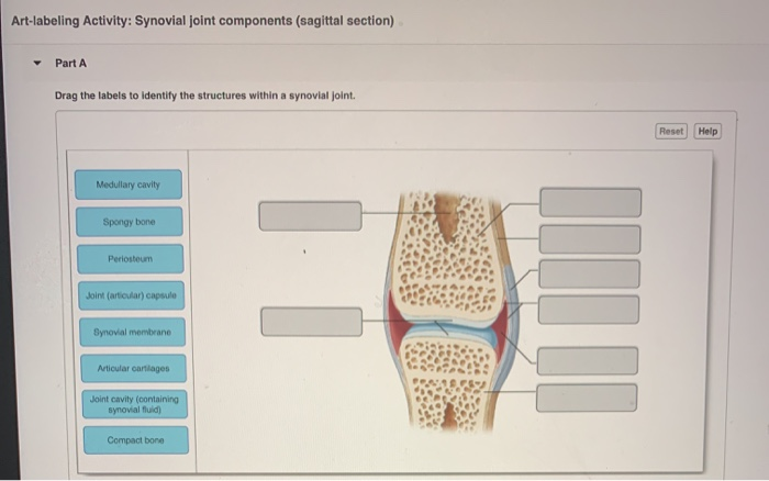
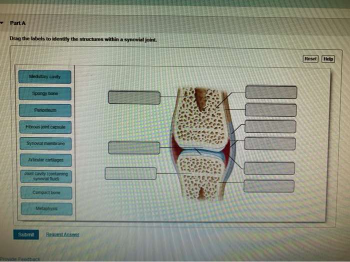
Art-labeling Activity: Structure of a Typical Synovial Joint
The Elbow Joint. Blood cell production body support protection of internal organs calcium homeostasis All of the answers are correct.

Synovial Joints Anatomy And Physiology I
The elbow is the joint connecting the upper arm to the forearm.
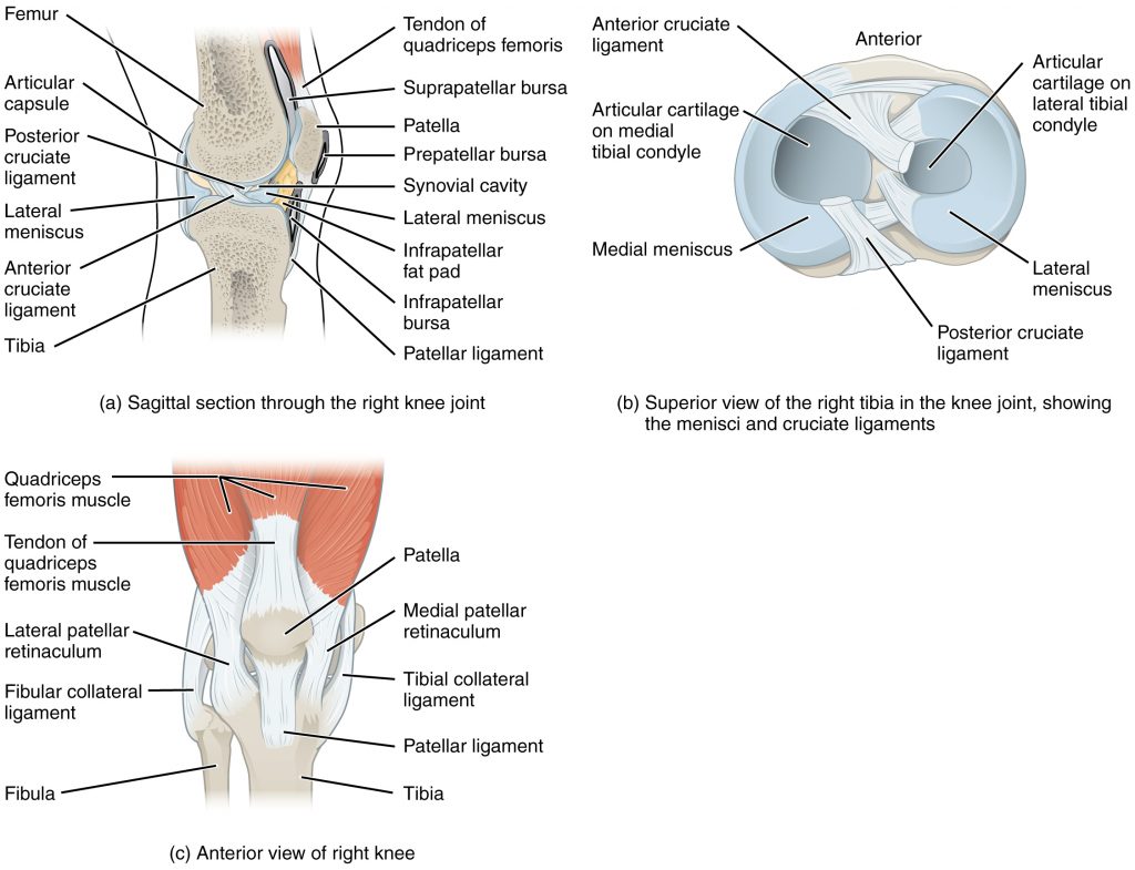
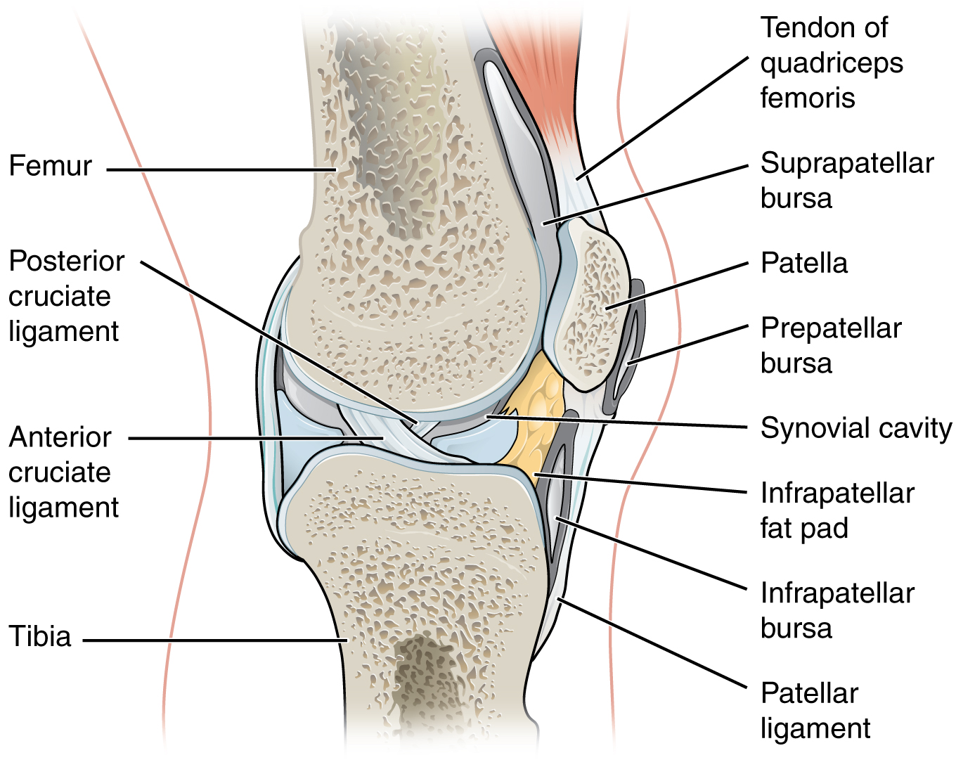
. The mandible is attached to the rest of the skull by a freely movable joint. Bursae are a part of synovial joints but tendon sheaths are not. The Knee Joint Drag the correct label to the appropriate structure of the knee joint.
Types of Synovial Joints. Human skull inferior view mandible removed Figure 512. B The hinge joint of the elbow works like a door hinge.
In this article we shall look at the anatomy of the elbow joint. Synovial Membrane inner- secretes produces synovial fluid for lubrication. Correct Spotlight Figure 82.
The structure of a long bone humerus of arm Figure 59. Key Structures of a Synovial Joint. View art labeling activity - vertebral anatomyjpg from BSC MISC at Miami Dade College Miami.
Structure of a hair and hair follicle Figure 410. Connective tissues consist of ligaments cartilage. - makes the synovial fluid and encloses.
It consists of two layers. Bone Markings Part 1. Correct Art-labeling Activity.
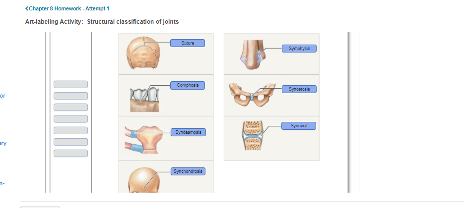
A Structural Classification of Synovial Joints Label the types of synovial joints. Structural features ligaments and associated tendons of the shoulder joint Art-labeling Activity. Learn vocabulary terms and more with flashcards games and other study tools.
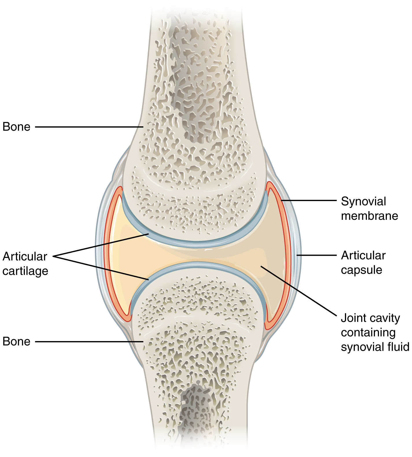
Flashcards Glossary Test Yourself. I articular capsule ii articular cartilage iii synovial fluid. The Structure of a Synovial Joint Art-labeling Activity.
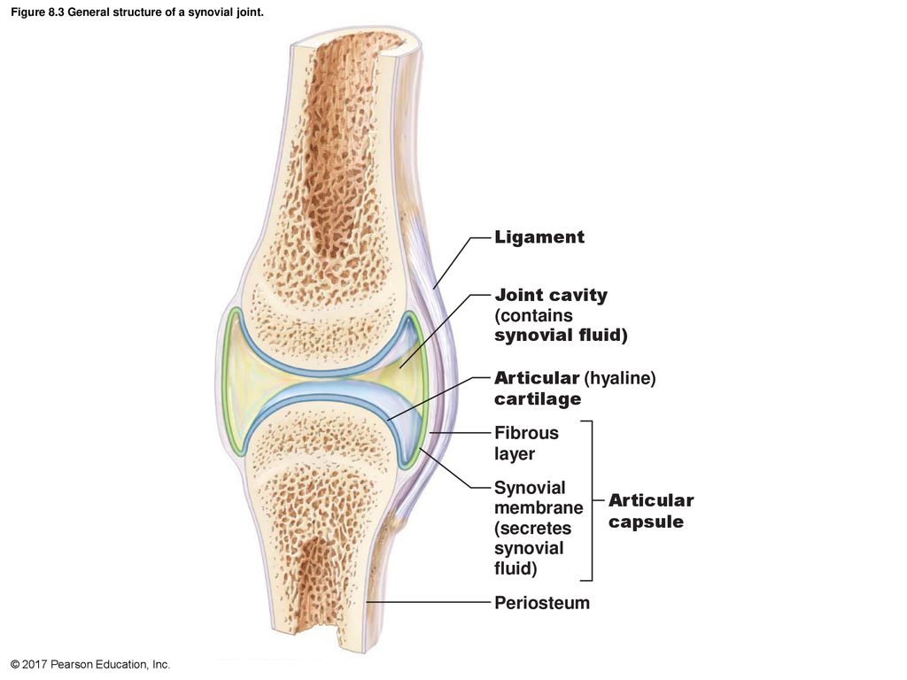
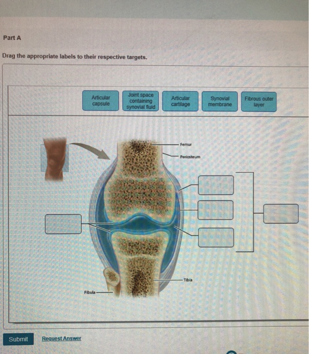
Art-labeling Activities Art-labeling Activity. Fibrous Capsule outer- provides joint stability. The articular capsule surrounds the joint and is continuous with the periosteum of articulating bones.
Structural classification of articulations - Anatomy Practice. To learn the bone markings. Label the bone markings.
The Shoulder Joint Art-labeling Activity. The six types of synovial joints allow the body to move in a variety of ways. No movement bursae reduce friction between adjacent structures but tendon sheaths do not.
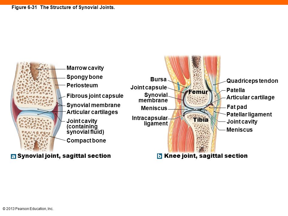
The Structure of a Synovial Joint. Its articulating surfaces movements stability and the clinical relevance. The right elbow joint medial view.
The three main features of a synovial joint are. Structure of a Synovial Joint - Anatomy Practice. Human skull anterior view.
The Right Elbow Joint Showing Stabilizing Ligaments Art-labeling Activity. Label the diagram of a typical synovial joint using the terms provided in the key and the appropriate leader lines. The role of joints and connective tissue.
Learn vocabulary terms and more with flashcards games and other study tools. 123131 are described in Tables 91 and 92 on p. Reset Help Fibula Lateral meniscus Anterior cruciate ligament Ligaments That Stabilize the Knee Joint Tbial collateral ligament Articular cartilage Fibular collateral ligament Patellar surface Tibia Patelar igament cut Posterior cruciate ligament Medial meniscus.
Slipping movements only Nonaxial movement. M no subject - stellarvore91gm x G anatomy abdominal quadrants - X Course Home X. A Pivot joints allow for rotation around an axis such as between the first and second cervical vertebrae which allows for side-to-side rotation of the head.
Activity 1 Identifying the Bones of the Skull The bones of the skull Figures 91910 pp. Drag the labels onto the diagram to identify the bone markings. A joint is a place where two or more bones meet and is also called an articulation.
Structure of a nail. Start studying The Structure of a Synovial Joint Sagittal section. Take the Chapter Practice Test to assess your progress and get your personalized study plan.
Movement in one plane Uniaxial movement. A typical synovial joint Figure 43. Muscles that move the hand and fingers anterior view middle layer Art-labeling Activity.
Movement in or around three planes Biaxial movement. As you read through this material identify each bone on an in-. - a sheet of cells that lines the joint cavity.
Human skull lateral view. Joint Motion Read through Spotlight Figure 82 and then complete the questions and activity below. Human skull superior view top of cranium removed Figure 511.
It is classed as a hinge-type synovial joint. Learn vocabulary terms and more with flashcards games and other study tools. Multiaxial movement movement in or around three.
Part A Drag the correct label to the appropriate location to identify the types of synovial joints. Skin structure Figure 48c. Interlocking fibrous joints called sutures.

Synovial Joints Anatomy And Physiology I
Solved Art Labeling Activity Synovial Joint Components Chegg Com
13 2 Musculoskeletal Basic Concepts Nursing Skills

Schematic Drawing Of An Idealised Synovial Joint Indicting Some Of The Download Scientific Diagram

Chapter 9 Question Set Flashcards Quizlet
Solved Art Labeling Activity Structure Of A Typical Chegg Com

The Role And Possible Mechanism Of Circrnas In Macrophage Cartilage Download Scientific Diagram
Answered Art Labeling Activity Structural Bartleby
Synovial Joints Anatomy And Physiology

Mastering A P Lab 8 Ex 11 Flashcards Quizlet

Ch 8 Art Labeling Activity Structure Of A Synovial Joint Anatomy Practice Flashcards Quizlet

Schematic Drawing Of An Idealised Synovial Joint Indicting Some Of The Download Scientific Diagram
Synovial Joints Anatomy And Physiology

Chapter 9 Question Set Flashcards Quizlet
2017 Pearson Education Inc Ppt Download
Solved Ch 09 Homework Joints Art Labeling Activity Chegg Com

My Lab And Mastering Chapter 8 Articulations Flashcards Quizlet
6 The Skeletal System Ppt Download

6 3 Bone Structure Anatomy Physiology


Comments
Post a Comment